
Delve into the intricate depths of neuroanatomy with our comprehensive Gross Anatomy of the Brain and Cranial Nerves Review Sheet. This meticulous guide unveils the structural intricacies of the brain and cranial nerves, empowering you with a profound understanding of their functions and clinical significance.
From the cerebral hemispheres to the cerebellum, from the brainstem to the cranial nerves, this review sheet meticulously dissects each component, providing a panoramic view of the brain’s anatomy and its pivotal role in our neurological functions.
Introduction
Understanding the gross anatomy of the brain and cranial nerves is crucial for comprehending the structure and function of the central nervous system. This review sheet provides a concise overview of the major anatomical features and their associated functions.
Cerebral Hemispheres
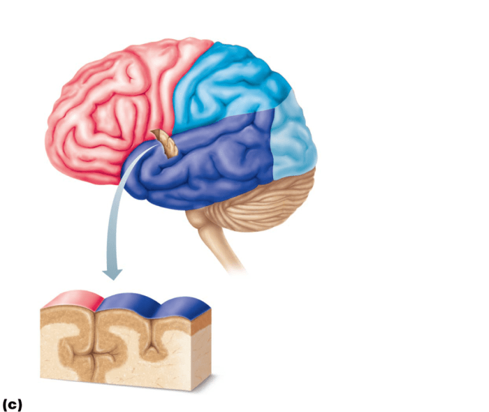
The cerebral hemispheres, the largest part of the brain, are divided into two hemispheres by the longitudinal fissure. Each hemisphere is further divided into lobes by sulci (grooves) and gyri (ridges).
- Frontal lobe:Responsible for higher-level cognitive functions, such as decision-making, problem-solving, and language production.
- Parietal lobe:Processes sensory information from the body and the environment.
- Temporal lobe:Involved in memory, language comprehension, and auditory processing.
- Occipital lobe:Processes visual information.
The corpus callosum, a bundle of nerve fibers, connects the two hemispheres and facilitates communication between them.
Brainstem
The brainstem, located inferior to the cerebral hemispheres, consists of the medulla oblongata, pons, and midbrain.
- Medulla oblongata:Controls vital functions such as breathing, heart rate, and blood pressure.
- Pons:Relays sensory and motor information between the cerebral hemispheres and the cerebellum.
- Midbrain:Contains nuclei involved in eye movements, hearing, and motor coordination.
The cranial nerve nuclei, located within the brainstem, give rise to the 12 pairs of cranial nerves.
Cerebellum

The cerebellum, located posterior to the brainstem, plays a crucial role in motor coordination and balance.
- External anatomy:Consists of two hemispheres with a central vermis and a series of transverse sulci and gyri.
- Functions:Receives sensory input from the spinal cord and other parts of the brain to coordinate muscle movements and maintain balance.
- Connections:Connected to the brainstem, cerebral hemispheres, and spinal cord.
Cranial Nerves

The 12 pairs of cranial nerves are responsible for transmitting sensory and motor information between the brain and various parts of the body.
| Cranial Nerve | Origin | Pathway | Function |
|---|---|---|---|
| I. Olfactory | Olfactory bulb | Through cribriform plate | Smell |
| II. Optic | Retina | Through optic foramen | Vision |
| III. Oculomotor | Midbrain | Through superior orbital fissure | Eye movements |
| IV. Trochlear | Midbrain | Through superior orbital fissure | Eye movements |
| V. Trigeminal | Midbrain and pons | Through trigeminal foramen | Sensory innervation of face |
| VI. Abducens | Pons | Through superior orbital fissure | Eye movements |
| VII. Facial | Pons | Through stylomastoid foramen | Motor innervation of facial muscles |
| VIII. Vestibulocochlear | Inner ear | Through internal acoustic meatus | Hearing and balance |
| IX. Glossopharyngeal | Medulla oblongata | Through jugular foramen | Sensory and motor innervation of tongue and pharynx |
| X. Vagus | Medulla oblongata | Through jugular foramen | Motor and sensory innervation of organs in thorax and abdomen |
| XI. Accessory | Medulla oblongata and spinal cord | Through jugular foramen | Motor innervation of neck and shoulder muscles |
| XII. Hypoglossal | Medulla oblongata | Through hypoglossal canal | Motor innervation of tongue muscles |
Cranial nerve injuries can lead to various neurological deficits, depending on the affected nerve.
Blood Supply to the Brain: Gross Anatomy Of The Brain And Cranial Nerves Review Sheet

The brain is supplied with blood by two major arteries, the internal carotid arteries and the vertebral arteries, which join to form the Circle of Willis.
- Circle of Willis:A network of arteries at the base of the brain that provides alternative routes for blood flow in case of blockages.
- Cerebrovascular accidents (strokes):Occur when the blood supply to the brain is interrupted, leading to tissue damage and neurological deficits.
Imaging of the Brain
Various imaging techniques are used to visualize the brain for diagnostic and treatment purposes.
- Magnetic resonance imaging (MRI):Uses magnetic fields and radio waves to create detailed images of the brain’s structure and function.
- Computed tomography (CT):Uses X-rays to create cross-sectional images of the brain.
- Limitations and risks:Brain imaging techniques may have limitations, such as the inability to detect certain types of brain abnormalities, and may involve risks, such as exposure to radiation.
FAQs
What is the purpose of the corpus callosum?
The corpus callosum facilitates communication between the left and right cerebral hemispheres, enabling the integration of sensory, motor, and cognitive functions.
How many pairs of cranial nerves are there?
There are 12 pairs of cranial nerves, each with a unique origin, pathway, and function.
What is the clinical significance of cranial nerve injuries?
Cranial nerve injuries can result in a wide range of neurological deficits, including sensory loss, motor weakness, and autonomic dysfunction.
